

Patella Dislocations and Treatment
by Steve Mora MD Knee Specialist
Taking care of patients who have anterior knee pain can be very challenging. Knee problems associated with the patella can be very challenging. The first in the evaluation of anterior knee pain is making the correct diagnosis. One common problem I see is patella instability.
Patella instability is a complex problem that has a wide range of contributing factors. Patients will often present with a history of having had a partial dislocation (subluxation) or a complete dislocation at some point in the past. In cases of complete dislocations the patella usually ends up on the lateral (outer) side of the knee.
 Picture of a laterally dislocated patella.
Often times it pops back in on its own upon extending the knee. After the acute event, the patient may continue to have anterior knee and/or persistent dislocations. After the dislocation patients will often times have persistent anterior knee pain and persistent episodes of subluxations. The persistent knee pain is often due to damage of the undersurface of the patella (articular cartilage) which occurs after the dislocation. In these cases the pain is due to both instability and traumatic chondromalacia of the patella.
 Damaged articular cartilage following a dislocation
The YouTube video link below shows a unstable patella in a high level Jiu Jitsu athlete prior to surgical treatment. I stabilized the patella successfully by reconstructing the Medial patellofemoral ligament (MPFL).
Click here for Youtube Link: Patella Instability
Persistent patella instability may be due to damage of the supporting ligament called the medial patella femoral ligament (MPFL) which helps to keep the patella centrally aligned. Some patients are predisposed to a dislocation. These patients may have been born with loose ligaments or a poorly formed trochlear groove (trochlea dysplasia). It may also be due to excessive angulation of the knee (extensor mechanism valgus). These problems have all been identified as risk factors in patients with recurrent patella dislocation.
Treatment of patella instability depends on the underlying problem. In cases where there is a mild imbalance of muscular forces on the patella, physical therapy may improve tracking. The goal of therapy is to evaluate the whole extremity starting at the hips. The patients who have anterior knee pain and instability oftentimes have hip muscular control problems, muscular weakness or core weakness. Addressing these issues are a main focus during nonoperative treatment. Patients who have a deficiency in the VMO strength may have increased pressure on the lateral patella, a.k.a. excessive lateral patella compress ion syndrome. Another important factor to look at during physical therapy is the lateral retinaculum and iliotibial band. These two structures can pull the patella laterally and also cause more stress on the lateral patella facet, as well as lead to instability. In cases where instability persists following nonoperative treatment, surgery may help to improve symptoms.
Surgery for patella instability depends on the contributing factors. For example, if there is abnormal extensor mechanism valgus a tibial tubercle osteotomy may be necessary. An osteotomy is a relatively big procedure which requires cutting and shifting the top of the shin bone in order to re-align the patella back into its groove. This requires a large incision, screws and a long period of crutch use.
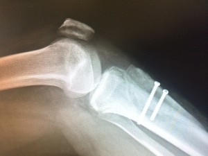 X-Ray showing a Fulkerson Tibial Tubercle Osteotomy. The shin bone has been cut and moved upward and inward.
In cases where there is a concomitantly stretched out MPFL, I may combine the tibial tubercle osteotomy with a ligament reconstruction. The ligament reconsctruction is called MPFL reconstruction. In some cases patients with instability may only need the MPFL ligament reconstruction and not the osteotomy. The ligament used to reconstruct the MPFL can be the patient’s own (autograft) or a cadaver graft (allograft). There are various types of tibial tubercle osteotomies but one of the most commonly used is called an anterior medial osteotomy a.k.a., a Fulkerson osteotomy. This type of osteotomy is not only good for instability but also very good for patients who have arthritis of the patella.
 X-Ray showing signs of a MPFL tear
Prior to proceeding with surgery, it is important I obtain plain x-rays and an MRI. Both are used to look at the position of the patella in relationship to the groove and the integrity of the patella cartilage. Prior to proceeding with the osteotomy, I typically do an arthroscopy to evaluate the knee for any loose cartilage or any other arthroscopically correctible abnormalities.
 MRI showing signs of a patella dislocation
In summary, the above surgical procedures for patella pain and instability can have favorable results in the right patient. For myself, the biggest challenge is making the correct diagnosis and identifying the contributing factors. As mentioned above, it is important to know whether the patella has under surface cartilage damage, determine whether or not abnormal bony anatomical issues exist and determine the integrity of the medial patellofemoral ligament. These surgical procedures, however, need to be done with extreme care. They do have a potential for significant complications. It is for this reason that I do everything possible to help my patients through their problem with nonoperative measures. The good news is that in cases where nonoperative treatment fails, there are surgical procedures with high success rate. The key once again is to accurately treat all the problems causing pain and instability.
I hope this information was helpful. If you would like further information you can contact me via my webpage or facebook.com/myorthodoc
About Steve A. Mora MD:

Dr. Mora is a native of Orange County. He graduated from Anaheim High School in Orange County CA. He completed his training at the UC Irvine where he finished in the top of his class with AOA Medical Society honors. He completed his Orthopedic Surgery training USC. He completed an extra year of training with a Sports Medicine, Cartilage, Shoulder, and Knee Fellowship at Santa Monica Orthopaedic and Sports Medical Group. He is currently practicing Orthopedic Surgery in Orange County.. Dr. Mora’s practice focus on sports related trauma, knee ligament and cartilage repair, shoulder rotator cuff and instability, hip arthroscopy and partial knee replacement. He sees athletes of all levels including professional soccer and UFC/MMA. He is team doctor for the Anaheim Bolts pro indoor soccer team and Foothill High School. Dr. Mora performs Cartilage transplantation (Genzyme), partial custom knee replacement, OATS, tibial osteotomies, meniscus transplant, ACL reconstruction, shoulder reconstruction, elbow arthroscopy, hip arthroscopy, platelet rich plasma and adult stem cell injections. Dr. Mora’s family heritage is Peruvian. He speaks fluent Spanish.
[embedyt]http://www.youtube.com/watch?v=Ic6qj-5zdgw&width=400&height=250[/embedyt]
 |
 |
 |
 |
|
Check out my Profile on Yelp
|
For more info become a Fan!
|
Dr. Mora’s Twitter Link: 4000+ Followers
|
My Professional LinkedIn Profile
|
![]()
Address:
Restore Orthopedics and Spine Center
112o W. La Veta Ave, Third Floor
Orange, CA 92868
Office: (714) 332-5498
Fax: (714) 941-9539
E-mail: smora@restoreorthopedics.com
Error: Contact form not found.
[slideshow_deploy id=’1142′]