
![]()
A Christmas Miracle- A Compound Fracture of the Tibia
Steve Mora MD
Orange County Fracture Specialist
Warning. Very graphic images below.
If you ask any Orthopedic surgeon about success stories they will tell you that there are too many to count. However if you ask them about cases that seemed to defy the expected natural course and result in an unbelievable or totally unexpected outcome there are always a couple that stand out. The cases that stand out are usually those which at first glance one would say there is no way that the outcome of this injury is going to be good. The story below is just that. Nothing short of a miracle.
The beautiful thing about the Orthopedic Surgery career is that we can help patients overcome difficult physical situations. These problems usually come about because of injury or degeneration. We treat patients who one day they were fine; going to work, providing for their families, enjoying a hobby and the next day their lives are literally turned upside down- they lose their jobs, lose medical insurance and lose their homes. As Orthopedic Surgeons we are witness to the fragile balance between a healthy happy life and a life of disability, destitute, and depression.
As Orthopedic Surgeons our training and experience allows us to help folks who suffer trauma. There are patients who overcome very difficult situations and surprise us with a good result. These patients always stand out in our minds. When we deal with patients who suffer a devastating injury we know that the odds are not in favor for the patient. The bad injuries are the ones that the surgeon will say early on to the patient things like “you’ll be lucky to walk again” or “you’ll be lucky to keep your leg”. In most cases we are right. But sometimes we are not.
I am personally a success story. Many people do not know this but the reason I went into Orthopedic Surgery was because my life was positively affected by many orthopedic surgeons after my bus accident. My legs were mangled. I had open fractures just like my patient below however it involved both of my legs. After many surgeries and complications I am able to do things that no one ever thought would be possible. I use my personal experience as daily reminders that sometimes there is hope when others think that there is none.
Here is my patient’s story. The final video of her is at the end of this article.
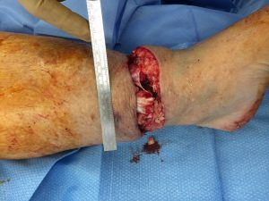
Two years ago, a couple of days before Christmas, while on ER call call, I was asked to see a 65 year old lady who had fallen off a ladder while cleaning out her rain gutters. She was alone working in her backyard on a ladder. She fell off the ladder and landed on her extended leg. Her lower leg bones, the tibia and fibula fractured. The force of the fall caused one of the sharp bone fragments to pierce the skin. This led to what is referred to as a compound or open fracture. As she fell, her body rotated causing the bone shard to slice through her lower leg skin and worsen the open fracture. It was a near-amputation meaning that the laceration was close to 360 degrees.
She was alone so she had to drag herself with her bleeding leg, bone jutting out of skin, to the phone. It was very difficult to hear how she struggled to get into her house to call 911. She told me this was one of the most difficult things she had ever had to do in her life. She could have easily bled to death but she did not. Here are some of the details.
I saw her in the ER and diagnosed her with the obvious open fracture of the tibia and arranged to operate on her right away.
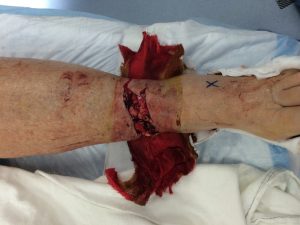
Warning. Graphic images below.
Pre op photo of the fracture:

The Christmas Miracle:


Once the swelling went down I removed the external fixation device and replaced it with an intra-medullary rod. External fixation devices work very well however they require extruding pins which can become infected. In this case I removed the external fixation device and replaced it with an intra-medullary rod. The rod continued to hold the fracture in line and allowed the patient to move without the cumbersome and heavy external frame. You can see the rod and a plate on the X-Ray. The fracture gaps started to fill in after about 3 months.


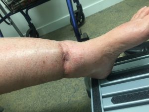

I did not see her for another year. Then one day she casually came in just before Christmas 2016 to see me not for the left ankle but because she sprained her opposite ankle being too active. It had been exactly 2 years from her fall off the ladder. She had recently returned from a cruise. I was so happy to see her. Here are the final X-Rays and her video.


-This patient was very fortunate that things worked out after her near amputation.
-If the sharp bone end had sliced just a couple more millimeters it would have damaged the artery and she would have probably lost her foot, but she did not.
-She could have lost enough blood at the time of injury that she could have succumbed from blood loss, but she did not.
-The open wound could have not healed and she could have required a complex and possible failed free muscle transplant over the soft tissue hole, but she did not.
-She could have developed a deep bone infection, but she did not.
-She could have developed a non healing fracture known as a non union, but she did not.
-She could have died from a hospital acquired pneumonia, but she did not.
-She could have lost major function of her ankle prohibiting her from walking without a walker and certainly not dancing, but she did not.
FINAL VIDEO OF OUR PATIENT
Main learning point here is that our bodies do in fact have a tremendous ability to heal. We are just starting to understand this healing potential especially as we explore regenerative healing therapies such as stem cells and platelets. I have been fortunately to have been part of the healing team for many other patients. As I think about these patients and their personalities they have something in common. They all had a positive attitude. They were fighters. They did not lose hope. It is this hope that helps me to also be a positive factor for patients who are suffering from injuries or degenerative musculoskeletal conditions.
Another learning point of my story: Do not climb up on ladders alone.
Please let me know if I can be of service.

![]()
Dr. Mora is a native of Orange County. He graduated from Anaheim High School in Orange County CA. He received his medical education at UC Irvine College of Medicine where he finished in the top of his class earning the coveted AOA Medical Society honors. He completed his Orthopedic Surgery training LAC+USC Medical Center and then did a additional Sports Medicine Fellowship at the Santa Monica Orthopaedic and Sports Medicine Group where focused on sports medicine, shoulder, knee, hip arthroscopy. He has published numerous book chapters on the topics of ACL injuries in soccer players, cartilage restoration, and athletic hip injuries. He is currently practicing Orthopedic Surgery in the City of Orange Orange County. He is a founding partner at Restore Orthopedics and Spine Center (www.restoreorthopedics.com). Dr. Mora’s practice focus is on sports related trauma, MMA injury treatment, arthroscopy of the shoulder, hip, knee and elbow, regenerative medicine (PRP and Stem Cells), partial and total knee replacement. He sees athletes of all levels including professional soccer and UFC/MMA patients. Dr. Mora’s family heritage is Peruvian. He speaks fluent Spanish.
 |
 |
 |
 |
|
Check out my Profile on Yelp
|
For more info become a Fan!
|
Dr. Mora’s Twitter Link: 4000+ Followers
|
My Professional LinkedIn Profile
|
Address:
Restore Orthopedics and Spine Center
112o W. La Veta Ave, Third Floor
Orange, CA 92868
Office: (714) 332-5498
Fax: (714) 941-9539
E-mail: smora@restoreorthopedics.com
Error: Contact form not found.
[slideshow_deploy id=’1142′]